Why should you X-ray the hips, elbows, and spine of your dog?
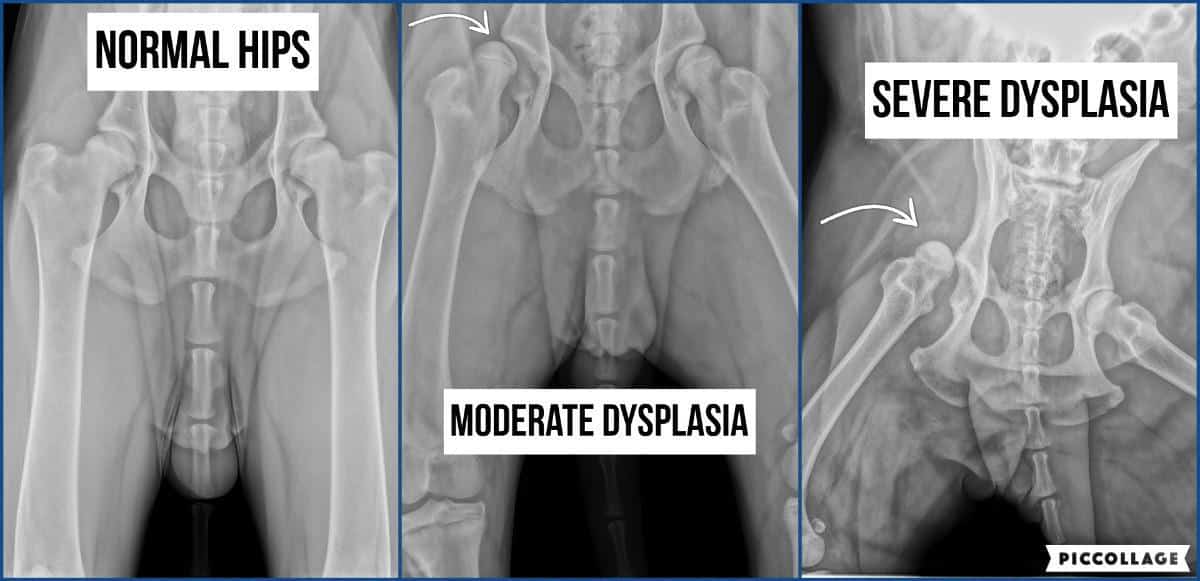
Hip dysplasia (HD) is not congenital because affected dogs are born with morphologically normal hips. HD is a developmental defect in the hip joint, where the femoral head (at the top of the thigh bone) does not fit together with the hip socket. This can cause abnormal mobility and wear and tear in the joint so the joint cartilage wears down and calcifications occur.

Dogs with HD can develop problems of varying degrees. Some dogs with HD experience no symptoms, while others may experience relatively severe pain and a reduced quality of life. Whether the dog will develop symptoms depends both on the degree of HD the dog has, but also on the dog’s construction, physical form, and area of use. Dogs with a healthy build that move efficiently and are kept in good physical shape (e.g. good muscles and kept rather thin) are less likely to develop symptoms than dogs with poor build and muscle mass, even if the dogs have the same degree of HD. The dog’s weight will for sure play a role, and should never have excessive weight.
Dogs with a mild degree of HD (grade C) will usually not develop any symptoms, possibly the symptoms will be mild and come late in life. Dogs with HD grade C will usually be able to live completely normal lives.
Dogs with moderate HD (grade D) will have a greater chance of developing symptoms. Many of the dogs in this group will do perfectly well, while others may develop symptoms that must be taken into account. In addition to the dog being in pain, the owner may find that the dog has a reduced utility value. In this group, there can be quite large individual and racial differences when it comes to symptoms.
Dogs with a severe degree of HD (grade E) run a much greater risk of developing symptoms than dogs with milder degrees. Dogs with grade E have the most severe symptoms, and they can appear early in life.
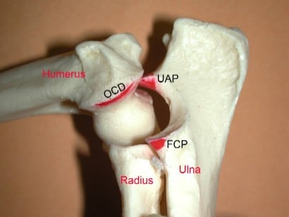
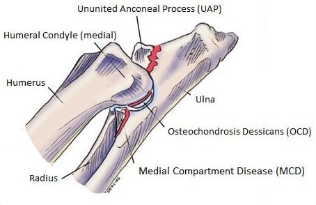
Elbow joint dysplasia (ED) is a collective term for various hereditary growth disorders that can occur in the elbow during the dog’s growth. These include fragmented processus coronoideus (FCP), osteochondrosis (OCD), ununited anconeal process (UAP), as well as non-specific osteoarthritis.


-
-
-
- Fracture of the ulna (FCP)
- Loose cartilage/osteochondrosis of the elbow joint (OCD)
- Loose bone fragments in the elbow joint (UAP)
- Non-specific osteoarthritis
-
-
If the veterinarian can detect any of these primary changes in the joint, the dog will be diagnosed with AD 3. If the dog has demonstrable calcifications (secondary changes) in the joint, the grade will be set from 1-3 depending on how severe the calcifications are.
The severity of symptoms also depends on the degree of maldevelopment and environmental factors. In the young dog, these usually occur as a result of cartilage damage, which is painful and can lead to lameness for periods or all the time throughout growing up. Dogs affected by elbow dysplasia often show signs from an early age, typically from 5 months on, but some may first be diagnosed after 4–6 years. Affected dogs develop a front limb lameness that typically worsens over weeks to months. Lameness is usually worse after exercise and typically never completely resolves with rest. Often both forelegs are affected, which can make detection of lameness difficult, as the gait is not asymmetric.
The cause of ED in dogs remains unclear. There are several theories as to the exact cause of the disease that include genetics, defects in cartilage growth, trauma, diet, and so on. It is most commonly suspected this is a multifactorial disease that causes growth disturbances.
HD and ED develop as the dog grows due to a combination of heredity and environment. A puppy is far from fully developed and especially the Rhodesian Ridgeback is a large breed that grows very quickly. The Airedale Terrier is also a medium to large size breed that grows quite quickly as well. It is therefore extremely important that the puppy gets the correct amount and the right type of feed. In addition, it should not be exposed to massive external influences such as playing unrestrained with very violent and large dogs or running for long periods on hard surfaces without the possibility of controlling the pace itself. Small puppies should be allowed to walk a lot on soft ground and move at their own pace, the very best is in rough and varying terrain such as forest. They are not fully grown until around the first sexual maturity, so before this time it is recommended to take account of growth. The dogs should NEVER have excess weight, and it’s better to keep the puppies and young dogs rather lean. The puppy should avoid walking on stairs until 4-5 months of age, and it should not be allowed to jump in and out of the car itself before it’s fully grown.

An early diagnosis of dysplasia through X-ray can help prevent further development of the disorder and reduce pain and discomfort for the animal. Early intervention can also provide the opportunity for treatment options such as physiotherapy, medication, or, in some cases, surgery. X-rays are therefore an important tool to ensure a good quality of life for our dogs.
The reading of HD/ED x-rays is also part of a screening program, where the aim is to reduce both the incidence and severity of HD/ED in the dog population. By giving individuals a HD/ED degree the occurrence of dysplasia in the various breeds and the various bloodlines and combinations can be mapped, as well as on an individual level.
Most puppy buyers and dog owners take measures to ensure that they are buying a dog where the probability of it developing severe HD and ED is small. You can get a good impression if the puppy’s parents and a large part of their siblings and extended family have been X-rayed and the results show a low or zero incidence of severe dysplasia. Although the degree of heritability is unknown, and the environment and upbringing are presumably of great importance, the genetic disposition will also have an impact. Thanks to previous puppy buyers having carried out these examinations on their dogs, the breeder of your next puppy can make well-considered choices regarding which dogs to breed on and which not. This is therefore a collective responsibility that you take on as a dog owner, and which can benefit yourself and others later.
HD/ED x-rays are recommended between 12-28 months and are done at a local vet who has an agreement with NKK (or another national kennel club if you live in another country) who reads the images and gives an official scoring. It is against the ethical basic rules for breeding and rearing in the Norwegian Kennel Club to breed from dogs with a strong degree of hip dysplasia or elbow dysplasia (HD grade E or ED grade 3).
Lumbosacral transitional vertebrae (LTV) is a congenital malformation in one of the vertebrae. The deformity occurs in the transition between the spinal segments (neck-chest, thoracic-lumbar, or lumbar-sacral), hence the name transitional vertebra. A transitional vertebra looks like a mixture of two types of vertebrae. Sometimes the transition vertebra is symmetrical, other times asymmetrical and can cause distortions in the back. A transitional vertebra usually has no clinical significance for the dog. In some cases, however, the malformation can cause the nerves at the bottom of the back to become pinched, which will cause symptoms such as back pain or limping.
It is not known with certainty how much stress this causes the dogs, but this is something that several ongoing research projects will try to uncover. A larger project has previously been carried out on the Ridgeback population in Finland, where it is now recommended to do X-rays for LTV at the same time as HD/ED, and it is mandatory for breeding. Dogs with proven LTV are recommended used in breeding if they are symptom-free, and bred to a partner with LTV0. Excluding these dogs with LTV1-4 from breeding when the heritability is not known is at the moment not recommended as it would involve the exclusion of a large proportion of the population. This in turn can lead to less genetic variation, which in turn can cause greater health challenges and more serious diseases to emerge due to increased inbreeding.
Transitional vertebra also occurs in humans and according to the Norwegian Medical Association, 5-10% of the population in Norway have this. Most people affected do not know they have it.
Excellent information about LTV can be read here: https://lumottu.net/?page_id=5874
LTV0: No abnormal findings
LTV1: Divided sacral crest (S1–S3) or some other mild abnormal structure
LTV2: Symmetrical lumbosacral vertebra
LTV3: Asymmetrical lumbosacral vertebra
LTV4: 6 or 8 lumbar vertebrae
Spondylosis Deformans (SP) is a degenerative spinal condition Rhodesian Ridgebacks also could be affected by. It is a condition that affects the vertebral bones of the spine and is characterized by the presence of bony spurs and bridges along the edges of the bones of the spine. A bony spur may develop in a single spot on the spine; more commonly, there will be multiple bone spurs in several different locations along the spine. Rhodesian Ridgebacks with an LTV appear to have a greater predisposition for spondylosis than the ones without it. Dogs with LTV1-LTV4 seem to have a significantly higher risk of spondylosis compared to dogs without, as 50% of dogs with spondylosis had also LTV (www.lumottu.fi). The exact cause of SP is unknown, but disease development might include repetitive trauma, instability, normal aging and wear and tear, and hereditary predisposition.
For a long time, spondylosis was considered an insignificant, asymptomatic aging change for dogs. However, it has been found that bone spurs and bridges formed in the spine can cause symptoms of varying degrees to the dog, such as stiffness, lameness, vague back pains, and reluctance to jump. The developing bone spurs can break or rub against each other, causing inflammatory pain in the area – sometimes the local symptoms are relieved when ossification progresses to a full bridge. The ventral bridge formation below the vertebrae stiffens the back, straining the adjacent intervertebral spaces. The rarer lateral spondylosis, which forms on the sides of the vertebrae, can press on the nerve roots and cause severe symptoms in the dog, such as urinary and fecal incontinence or paralysis symptoms.
SP0: Free, no abnormal findings
SP1: Mild, < 3 mm spurs in ≤ 4 intervertebral spaces or > 3 mm spurs in ≤ 3 intervertebral spaces or a bone island in ≤ 2 intervertebral spaces.
SP2: Evident, bone bridges (complete or incomplete) between ≤ 2 intervertebral spaces and/or large bone islands in ≤ 2 intervertebral spaces.
SP3: Moderate, bone bridges (complete or incomplete) and/or large bone islands between 3–7 intervertebral spaces.
SP4: Severe, more severe changes than above.
Vertebral anomaly (VA) occurs in several dog breeds and is an umbrella term describing a variety of different birth defects of the vertebral column such as butterfly vertebrae (partial or complete failure of formation of the ventral and central portions of the vertebral body), hemivertebrae (failure to form one sagittal half of the vertebrae, including the centrum and neural arch), wedge-shaped vertebrae (incomplete failure to form one sagittal half of the vertebrae with a variable degree of unilateral hypoplasia), transitional vertebrae (vertebra showing features of two adjacent segments of the vertebral column e.g. thoracic and lumbar), articular process dysplasia (aplasia or hypoplasia of the cranial or caudal articular process).
VA0: Normal, no abnormal findings
VA1: Mild, 1–2 malformed vertebrae (e.g. transitional T13 or L1)
VA2: Evident, 3–4 malformed vertebrae
VA3: Moderate, 5–9 malformed vertebrae
VA4: Severe, 10 or more malformed vertebrae
LTV/VA/SP screening in dogs is not yet mandatory in Norway, and I will at this time not insist that you do it. But if you want to know more about the status of your dog’s spine, I would recommend that you ask for lateral images covering at least the thoracic and lumbar vertebrae to be taken at the same time as HD/ED x-ray, for example, two to three images covering C7-L1 and T13-Cd1 (gives an evaluation of LTV and SP). If you also want to have a VA screening you also have to as one image covering C1-C7.
There is currently no official screening program in Norway, but a veterinary evaluation regarding LTV/VA/SP can be done by sending pictures to INCOC which is a reporting service of screening radiographs for dog owners. INCOC is owned by Anu Lappalainen DVM, Ph.D. and Vilma Reunanen DVM, both Finnish Kennel Club scrutineers for HD, ED, shoulder OC, spinal (IDD, LVT, SP, VA), and INC radiographs.
The dog must be at least 12 months old when x-rayed, and then the dog receives a statement that includes congenital changes in the spine (i.e. LTV and VA). A dog that has turned 24 months old at the time of the x-ray can also be given a statement that covers developmental changes in the back (SP). I usually x-ray my dogs before 24 months of age, which means that I take additional photos of the spine after 24 months of age to get the SP statement in addition.
Sources
- www.forthvet.com.au/
- www.fitzpatrickreferrals.co.uk
- www.acvs.org/
- www.vcahospitals.com/
- www.ofa.org
- www.retrieverklubben.no/
- www.nkk.no/
- www.lumottu.net/
- www.kennelliitto.fi

